How Much Does an FDG PET CT Scan Cost?
MIC uses FDG PET CT imaging in several applications: heart (cardiac) imaging, cancer (oncology) imaging, and brain (neurology) imaging. Currently, the Alberta Health Care Insurance Plan (AHCIP) only covers the cost of FDG PET CT imaging in cardiac applications when performed in a community-based clinic outside the hospital.
The cost of FDG PET CT imaging in oncology or neurology applications is as follows:
| PET CT Imaging Exam | Cost |
|---|---|
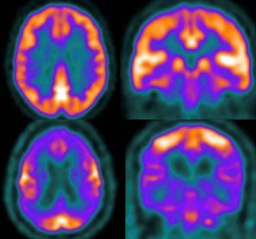
| FDG PET CT – Brain Scan | $999.00 |
| FDG PET CT – Standard Whole Body | $1,099.00 |
| FDG PET CT – Extended Whole Body | $1,199.00 |
| FDG PET CT – Head and Neck Package* | $1,499.00 |
| Diagnostic Enhanced CT Scan | $499.00 |
Please note:
- Our dual-trained radiologist and nuclear medicine physicians will protocol all requisitions to ensure an accurate price estimate.
- The price is based on the imaging application, relevant tracer, area of interest (brain, head, neck, whole body, etc.), and need for a combined diagnostic enhanced CT.
- If a combined PET and diagnostic enhanced CT is required, the CT component will be $499.
- There is no cost for FDG PET CT imaging in cardiac applications for patients with a valid Alberta Health Care card or an out-of-province health care card (except Quebec).
- *FDG PET CT – Head and Neck Package includes a standard whole-body scan with separate, dedicated, high-resolution neck imaging.*
Patients are encouraged to talk to their employer or a Benefits Specialist before booking their exam to see if they qualify for extended coverage. Some extended benefits programs or health spending accounts may cover a portion or possibly the entire exam fee.
Hotel Partnerships
We are also happy to help travelling patients take advantage of price discounts with two local hotels!
Patients flying into Edmonton should consider taking advantage of our partnership with the Renaissance Edmonton Airport Hotel.
Patients driving into Edmonton may prefer to stay at the Delta Hotels Edmonton South Conference Centre, which is conveniently located near our Century Park clinic, where PET CT imaging is offered.
Be sure to ask our booking team for detailed instructions on how to save money if you are considering travelling for your PET CT exam.
FDG PET CT FAQs
The MIC PET requisition provides ordering physicians with a number of opportunities to specify what specific areas they would like scanned. If there is ongoing uncertainty, the radiologist will contact the ordering physician to ensure the scan is optimized to address the clinical question.
Each of these PET scans provides complementary information to aid the clinician in making the optimal personalized treatment decision for each individual.
Bois, J., Muser, D., Chareonthaitawee, P. (2019). PET/CT Evaluation of Cardiac Sarcoidosis. https://med.emory.edu/departments/radiology/clinical_divisions/nuclear-medicine/documents/2020/nuclear-cardiology/pet.ct-evaluation-of-cardiac-sarcoidosis.pdf
Berti, V., Pupi, A., Mosconi, L. (2011, June). PET/CT in diagnosis of movement disorders. https://pubmed.ncbi.nlm.nih.gov/21718327/
Fazio, P., Varrone, A. (2019, March). FDG-PET/CT in Movement Disorders. https://doi.org/10.1007/978-3-030-01523-7_6
Goel, R., Moore, W., Sumer, B., Khan, S., Sher, D., Subramaniam, R. (2017, July). Clinical Practice in PET/CT for the Management of Head and Neck Squamous Cell Cancer. https://www.ajronline.org/doi/10.2214/AJR.17.18301
Greenspan, B. (2017, December). Role of PET/CT for precision medicine in lung cancer: perspective of the Society of Nuclear Medicine and Molecular Imaging. https://pmc.ncbi.nlm.nih.gov/articles/PMC5709134/
Hofman, M., Hicks, R. (2016, October). How we read Oncologic FDG PET/CT. https://doi.org/10.1186/s40644-016-0091-3
Khalaf, S., Al-Mallah, M. (2020, April). Fluorodeoxyglucose Applications in Cardiac PET: Viability, Inflammation, Infection, and Beyond. https://pmc.ncbi.nlm.nih.gov/articles/PMC7350806/
Litt, H., Kwon, D., Velazquez, A. (2023, July). FDG PET Scans in Cancer Care. https://jamanetwork.com/journals/jamaoncology/fullarticle/2807033
Marcus, C., Paidpally, V., Antoniou, A., Zaheer, A., Wahl, R., Subramaniam, R. (2015, February). FDG PET/CT and Lung Cancer: Value of Fourth and Subsequent Posttherapy Follow-up Scans for Patient Management. https://jnm.snmjournals.org/content/56/2/204
Ng, S., Lau, H., Raynor, W., Werner T., Saboury, B., Revheim, M., Alavi, A. (2022, August). Diagnosis and evaluation of cardiac sarcoidosis with FDG PET/CT and PET/MR. https://jnm.snmjournals.org/content/63/supplement_2/2641
Skali, H., Schulman A., Dorbala, S. (2013, April). FDG PET/CT for the Assessment of Myocardial Sarcoidosis. https://pmc.ncbi.nlm.nih.gov/articles/PMC4009625/
Society of Nuclear Medicine and Molecular Imaging. (2023). Fact Sheet: Molecular Imaging and Colorectal Cancer. https://snmmi.org/Patients/Patients/Fact-Sheets/Molecular-Imaging-and-Colorectal-Cancer.aspx
Waxman, A., Herholz, K., Lewis, D., Herscovitch, P., Minoshima, S., Ichise, M., Drzezga, A., Devous, M., Mountz, J. (2009, February). Society of Nuclear Medicine Procedure Guideline for FDG PET Brain Imaging. https://snmmi.org/common/Uploaded%20files/Web/Centers/PET%20Center%20of%20Excellence/SNM%20Procedure%20Guideline%20for%20FDG%20PET%20Brain%20Imaging.pdf


