For more patient information regarding Breast Biopsies
Breast Biopsy Exam Prep
Find out when you book
What is a breast biopsy?
A breast biopsy is a procedure used to find out more about lumps or abnormalities that are detected by physical examination, mammography or other imaging studies. Using image guidance is the most accurate way to do biopsies. MIC radiologists perform breast biopsies using image-guidance. These types of biopsies are not intended to remove the growth, but to obtain a small sample for further analysis.
During a breast biopsy, your radiologist will take a tiny tissue sample from a suspicious area in your breast and send it to a laboratory to determine whether the growth is benign or cancerous. There is little or no noticeable scar after healing.
Your practitioner and radiologist will determine the best method to perform your biopsy. The exam may be different depending on which of the options they select. All procedures are performed with local freezing.
- In a Cyst Aspiration the radiologist uses ultrasound to guide a very thin needle and syringe to the abnormality in your breast to remove fluid, usually to relieve symptoms such as pain.
- In an Ultrasound-Guided Core Biopsy the radiologist uses ultrasound to guide a special hollow needle to the abnormality in your breast and take a tiny cylinder-shaped (core) sample of tissue.
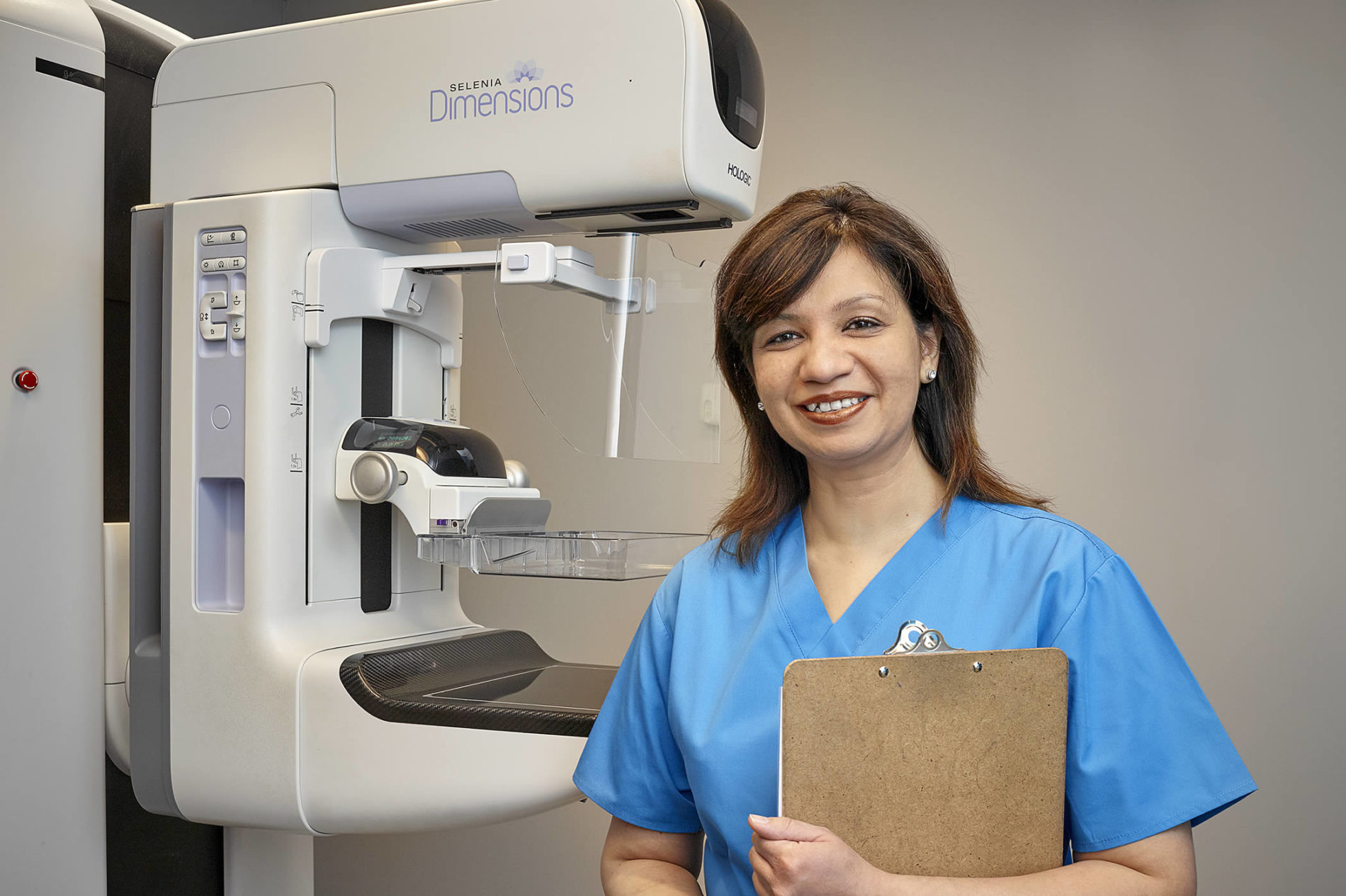
- In a Tomosynthesis or Stereotactic Breast Biopsy the radiologist uses a special mammography machine to guide a needle to the abnormality in your breast to take a small tissue sample.
- In a Wire Localization procedure, the radiologist inserts a fine wire to mark the exact location of the area of concern before a surgeon removes the lump or abnormal tissue.
What to expect
- When you arrive for your appointment, you will be asked to remove your clothing from the waist up and change into a gown.
- Your technologist will help position you on the table for your exam.
- Your radiologist will administer local freezing and use ultrasound to guide a thin needle into the correct location.
- The radiologist will confirm the correct location by viewing the needle from multiple angles and will let you know when the procedure begins.
- After the procedure is over, your technologist will go over detailed post-procedure care instructions with you.
- MIC will send the tissue samples to the lab for analysis. You will hear from your practitioner regarding results.
*Diabetic patients who use a glucose monitoring device should check with their manufacturer to see if they recommend removing their device before low-dose radiation exposure.
